
Our abdominal region, is a complex network of organs that play crucial roles in digestion, metabolism, and overall health. Understanding the nuances of this vital region is paramount for comprehensive healthcare. Enter abdominal sonar, a revolutionary imaging technique that peers into the depths of your abdomen without invasive. Abdominal sonar is a non-invasive imaging technique that utilizes sound waves to create detailed images of the internal structures of the abdomen. This non-invasive imaging technique utilizes sound waves to unveil the mysteries within, offering detailed insights into the structure and function of the organs nestled in our midsection.
This diagnostic tool provides valuable insights for healthcare professionals, helping them assess and diagnose various conditions affecting the liver, gallbladder, kidneys, pancreas, and other abdominal organs. Understanding your tummy goes beyond surface perceptions, and abdominal sonar emerges as a window into the intricate workings of an often-underestimated part of the human body. Let’s delve into the basics of abdominal sonar, exploring how it works, its applications, and the vital information it can unveil about your tummy health.
What is Abdominal Sonar?
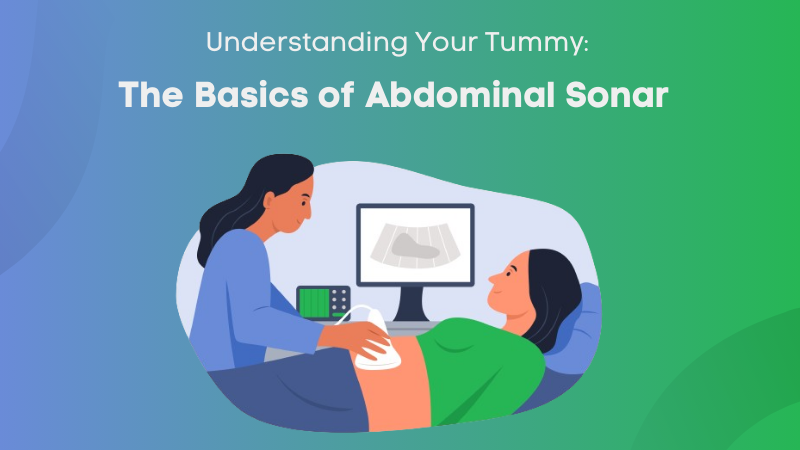
Abdominal sonar, also known as abdominal ultrasound, is a non-invasive medical imaging technique designed to visualize the internal structures of the abdomen using high-frequency sound waves. Employing a transducer wand that emits these sound waves into the body, the technology captures the echoes as they bounce back from various tissues. This process allows for the creation of real-time images on a monitor, offering detailed views of abdominal organs like the liver, gallbladder, pancreas, kidneys, and spleen.
Unlike imaging methods that use ionizing radiation, abdominal sonar is considered safe and is commonly utilized for a range of diagnostic purposes. From monitoring pregnancies to identifying abnormalities and assessing the health and function of abdominal organs. This dynamic imaging tool has become an integral part of modern healthcare, providing valuable insights into the complexities of the abdominal region without the need for invasive procedures.
How Abdominal Sonar Works?
Abdominal sonar begins with a transducer wand, a crucial component of the sonar system, emitting high-frequency sound waves. These waves penetrate the body and bounce back when they encounter different tissues, reflecting unique echoes. The transducer not only emits but also receives these echoes. A computer processes the echoes, translating them into real-time images displayed on a monitor. The resulting images showcase the varying shades and textures of different tissues, allowing healthcare professionals to discern the contours and functioning of organs.
Notably, the real-time imaging capability of abdominal sonar enables dynamic observation, making it an invaluable tool for diagnosing conditions and guiding medical interventions with precision. The non-invasive nature of this technology, coupled with its ability to capture immediate, live images, distinguishes abdominal sonar as a versatile and indispensable diagnostic tool in the realm of medical imaging.
What to Expect During An Abdominal Sonar
During an abdominal sonar, patients can expect a straightforward and minimally invasive procedure. Here’s a breakdown of what to expect:

1. Preparation
Depending on the specific purpose of the sonar, your healthcare provider might give you specific instructions. In some cases, you might be asked to fast for a few hours before the procedure, especially if the focus is on the upper abdominal organs. When you fast for a few hours before the procedure, your stomach is less likely to contain undigested food, providing a clearer acoustic window for the sound waves to penetrate and produce accurate images. Typically, healthcare providers may ask patients to abstain from consuming food and liquids for about 6 to 8 hours before the abdominal sonar.
2. Examination Room
Abdominal Sonar typically performed in a dimly lit room to enhance visibility on the monitor. Dimly lit room create an atmosphere conducive to the visualization of images on the monitor. The subdued lighting helps enhance the contrast on the screen, allowing healthcare professionals to obtain clearer and more detailed images of the abdominal organs. This careful control of the lighting conditions is a deliberate measure to ensure the accuracy and effectiveness of the sonar examination, providing the medical team with the best possible insights into the health and functioning of your abdominal structures.
3. Positioning
As you lie down on the examination table, the next step in the abdominal sonar procedure involves the exposure of the specific area of your abdomen that needs examination. This is necessary for the effective transmission of sound waves from the transducer wand to the internal structures. The healthcare professional will apply a clear, water-based gel on the area. The gel serves multiple purposes as it helps eliminate air pockets between the transducer and the skin, ensuring uninterrupted sound wave transmission. The gel application is a standard and essential part of the sonar process, contributing to the overall success of the examination.
4. Transducer Application
The transducer wand is responsible for emitting high-frequency sound waves into the body and receiving the echoes that bounce back from internal structures. The transducer is gently placed on the gel-covered area of your abdomen, and the healthcare technician skillfully moves it over the skin in a controlled manner. This movement allows the transducer to capture real-time images of the internal structures, including organs like the liver, gallbladder, pancreas, kidneys, and others. The technician may adjust the angle and position of the transducer to obtain different views, ensuring a thorough examination of the targeted area.
5. Real-Time Imaging
During the abdominal sonar, the transducer captures the echoes generated by the sound waves as they bounce back from the internal structures of your abdomen. These echoes are swiftly processed by a computer in real-time, creating a dynamic and detailed set of images. The real-time aspect of the procedure is particularly valuable, as it allows both you and the healthcare professional to observe the movement and functionality of the organs as they are being imaged. This real-time interaction between the sonographer and the images on the monitor is a key element in the diagnostic process, ensuring a meticulous examination of your abdominal health.
6. Duration
The procedure is typically brief, lasting approximately thirty minutes, making it a convenient diagnostic tool for various abdominal health assessments. Importantly, the process is painless, alleviating concerns related to discomfort or post-procedural recovery. As the examination concludes, the healthcare professional will swiftly and easily wipe off the gel applied to your skin during the procedure. This step ensures your comfort and allows you to resume your daily activities immediately.
7. Post-Procedure Information
Your healthcare provider may offer you some initial insights into the findings. In some cases, they might discuss preliminary results immediately after the procedure, providing you with a brief overview of what was observed during the examination. This immediate feedback can offer some reassurance and initial information about your abdominal health. However, it’s important to note that a comprehensive analysis of the images may require more time. In many instances, healthcare professionals prefer to conduct a thorough review of the sonar images before providing a detailed interpretation.
Conclusion
Abdominal sonar is a valuable tool in the realm of diagnostic imaging, providing detailed insights into the health and functioning of the abdominal organs. This non-invasive imaging technique has proven to be a valuable diagnostic tool, offering real-time insights into the functioning of vital organs. As we navigate the dynamic landscapes within our abdomens, it is essential to emphasize the importance of regular check-ups and consultations with healthcare professionals. A skilled gastroenterologist in Pretoria can provide specialized expertise in digestive health, offering personalized insights and recommendations based on abdominal sonar findings. By partnering with a healthcare expert, you can embark on a journey towards proactive abdominal health management.