

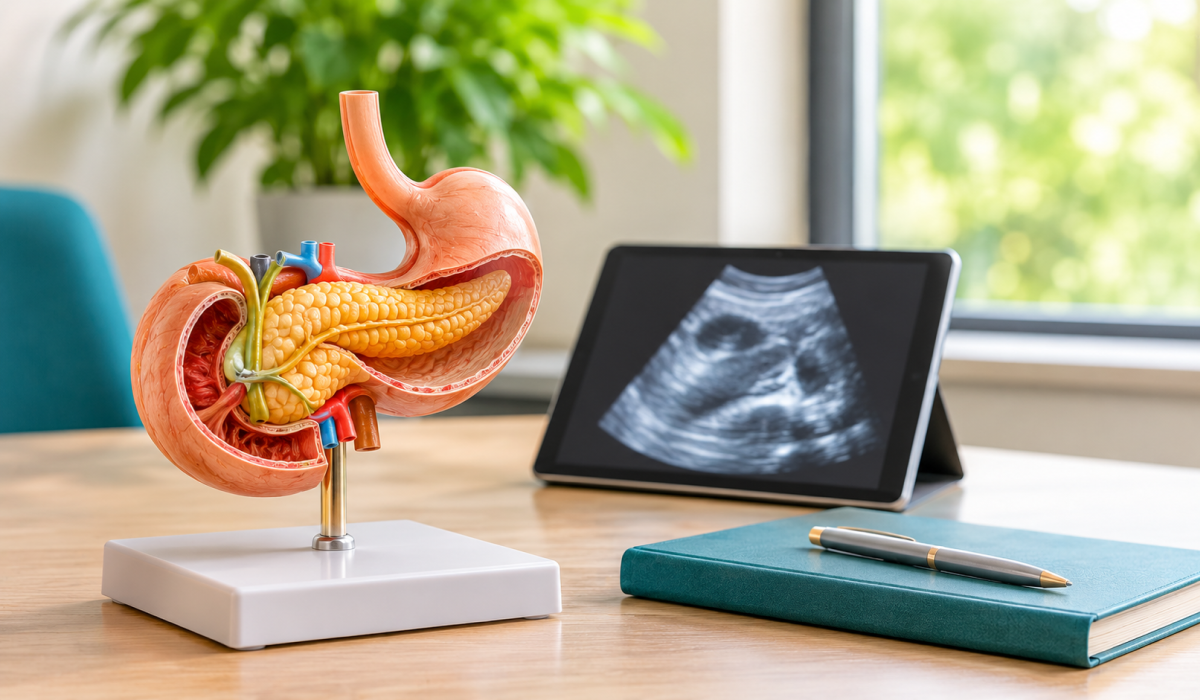
The pancreas is one of the most important organs in your body that most people never think about. It sits deep in the upper abdomen, tucked behind the stomach, and it does two vital jobs: it makes the digestive enzymes that break down food, and it produces insulin and other hormones that control blood sugar.
Because it’s so deeply placed, it’s also one of the hardest organs to see clearly.
That’s why, when a doctor needs a closer look, they often turn to endoscopic ultrasound for pancreas evaluation. It’s a procedure that combines endoscopy with ultrasound imaging, which lets a gastroenterologist examine the pancreas from just a few millimetres away instead of from outside the body.
This guide explains how it works, what it can find, what the experience is like, and where its limits lie, so you can walk into the conversation with your doctor better informed.
What Is an Endoscopic Ultrasound of the Pancreas?
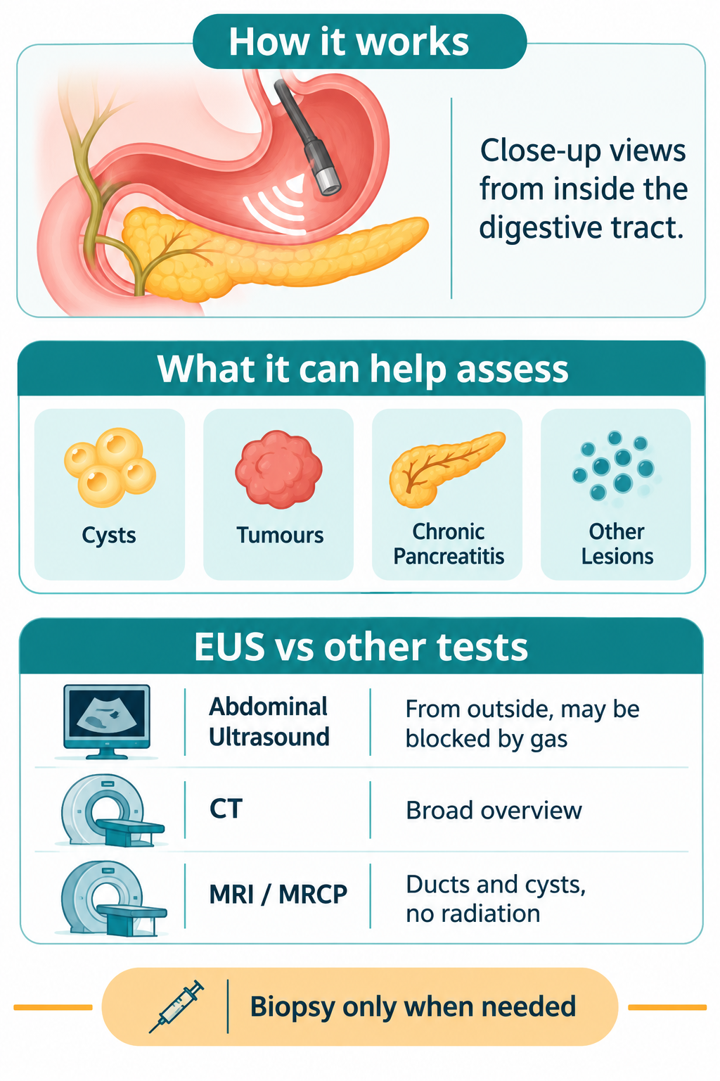
An endoscopic ultrasound, usually shortened to EUS, is a procedure that combines two tools in one. An endoscope is a thin, flexible tube with a light and a camera.
At its tip sits a small ultrasound probe, which sends out sound waves and uses the echoes to build up images. As Mayo Clinic explains, the ultrasound device at the end of the tube generates images of the digestive tract and nearby organs and tissues.
When EUS is used for the pancreas, it’s sometimes called pancreatic endoscopic ultrasound, or EUS for pancreas.
The idea is simple. Because the pancreas lies right behind the stomach and next to the first part of the small intestine, the probe can sit very close to it from inside the digestive tract.
That closeness gives a clear, detailed picture of the pancreatic tissue, the pancreatic duct, and the structures around it.
It differs from a standard abdominal ultrasound in an important way. With an abdominal ultrasound, the probe is pressed against your skin, and the sound waves have to pass through skin, fat, muscle, and often gas in the bowel before they reach the pancreas.
That can blur the picture, and in some people the pancreas can’t be seen well at all. EUS removes most of those obstacles.
Why Is Endoscopic Ultrasound Used to Examine the Pancreas?
A gastroenterologist may recommend EUS when they need more information than other tests have provided. Common situations include:
- A scan such as a CT or MRI has shown something unclear, such as a small spot, a cyst, or a possible mass
- You have unexplained upper abdominal pain, repeated episodes of pancreatitis, or unexplained weight loss, and the cause hasn’t been found
- A doctor wants to look closely at the pancreatic duct or the tissue around it
- A suspicious area needs to be sampled for laboratory testing
EUS can look at pancreatic tissue and nearby structures at close range, including lymph nodes and blood vessels. It’s particularly useful for investigating unclear findings from earlier imaging, and it can help in diagnosing pancreatic abnormalities that are small or hard to see from outside the body.
The results then help your care team decide what to do next, whether that’s simply monitoring, further testing, or planning treatment.
It’s worth being clear on one point. EUS is a diagnostic tool, and it’s usually not a first test. It tends to come after a conversation, an examination, and often other imaging, when a more detailed look is needed.
What Pancreatic Conditions Can an Endoscopic Ultrasound Detect?
EUS can help assess a range of conditions. Here are the main ones.
Pancreatic Cysts
Pancreatic cysts are fluid-filled sacs in or on the pancreas. They’re surprisingly common, and many turn out to be harmless. Some types, however, can carry a small risk of becoming cancerous over time, which is why they’re looked at carefully.
EUS lets the doctor examine a cyst’s size, shape, wall, and internal features, such as dividing walls or small solid nodules, and see whether it connects to the pancreatic duct.
In some cases, a small amount of fluid can be drawn out with a fine needle and tested. Guidelines commonly recommend EUS for cysts that cause symptoms, are larger, or show features of concern, though the right approach always depends on the individual.
Pancreatic Tumours and Cancer
EUS can help identify suspicious growths in the pancreas, assess their size and location, and show how close they are to nearby blood vessels and lymph nodes.
That information matters when doctors plan treatment. It’s also one of the ways a sample of tissue can be obtained from a suspicious area, which is usually needed to confirm a diagnosis.
It’s important to be honest about what EUS can and can’t do. It’s a very sensitive way of looking at the pancreas, but no test is perfect, and a diagnosis of pancreatic cancer is never made from one image alone.
It’s usually based on a combination of imaging, tissue results, and the whole clinical picture. EUS is also not a routine screening test for people without symptoms or risk factors.
Chronic Pancreatitis and Pancreatic Inflammation
Chronic pancreatitis is long-term inflammation of the pancreas, often causing pain and digestive problems.
EUS can show changes in the pancreatic tissue and ducts that point towards it, such as altered texture, small calcium deposits, or irregular or widened ducts. It’s one of the tools used when the diagnosis isn’t clear from symptoms and other scans.
Pancreatic Lesions and Other Abnormalities
“Lesion” simply means an unusual area in the tissue. EUS helps assess these, and it can reveal small abnormalities that other scans miss.
Sometimes, though, the picture is still uncertain, and further tests, such as other scans, tissue sampling, or monitoring over time, are needed to reach a clear answer. That’s a normal part of the process, not a sign that something has gone wrong.
How Does Endoscopic Ultrasound Work for the Pancreas?
If you’re wondering how the procedure unfolds, it helps to think of it in three steps.
Inserting the Endoscope
You’ll usually be sedated, and a thin, flexible endoscope is gently passed through your mouth, down the oesophagus, and into the stomach and the first part of the small intestine.
If you’ve had a gastroscopy before, the start of the procedure will feel familiar, but the EUS instrument has the added ultrasound probe at its tip.
Using Ultrasound to Examine the Pancreas
From inside the stomach and the first part of the small intestine, the probe sends out sound waves and receives the echoes, creating images of the pancreas and surrounding structures.
Different parts of the pancreas are best seen from different positions: the body and tail from the stomach, and the head from the duodenum. The doctor moves the probe gently to look at the pancreas from several angles.
Capturing Detailed Images
The images appear on a screen in real time, and the doctor can also save them. Together they show the texture of the pancreatic tissue, the shape and width of the duct, and any cysts, lumps, or enlarged lymph nodes.
Because the images are so detailed, they help the doctor decide whether something needs to be sampled, monitored, or followed up with more tests.
What Is an EUS-Guided Biopsy of the Pancreas?
If something suspicious is seen, your doctor may take a sample of cells or tissue during the same procedure, using the ultrasound image as a guide. This is called an EUS-guided biopsy. A very thin needle is passed through the endoscope and into the area of concern, watching on the screen to guide it precisely.
There are two main types:
- Fine needle aspiration (FNA) draws out cells or fluid, which are then examined under a microscope (a test called cytology).
- Fine needle biopsy (FNB) takes a small core of tissue, which keeps the structure of the tissue intact so the pathologist can study it in more detail.
Either type may be used, depending on what’s being sampled and what information the doctor needs. The samples are sent to a laboratory, and the results usually take several days.
A biopsy is not part of every EUS. Many examinations are purely imaging, and a biopsy is only done when there’s a clear reason for it.
How to Prepare for a Pancreatic Endoscopic Ultrasound
Preparation for EUS is similar to preparation for other upper endoscopy procedures, but you should always follow the specific instructions you’re given.
- Fasting. You’ll usually be asked not to eat or drink for several hours beforehand, often six to eight, so your stomach is empty.
- Medicines. Tell your doctor about everything you take, especially blood thinners, diabetes medication, and supplements. Some may need to be adjusted, and you shouldn’t change anything on your own.
- Allergies and health conditions. Mention any allergies, heart or lung problems, and previous reactions to sedation or anaesthetic.
- Transport. If sedation is used, you’ll need someone to take you home, and you shouldn’t drive for the rest of the day.
- Instructions. Follow the written instructions from your medical team, and ask if anything is unclear.
If a biopsy is likely, your doctor may also ask about bleeding problems and may ask you to pause blood-thinning medication beforehand. That decision is individual, which is why it needs to be discussed in advance.
What Happens During an Endoscopic Ultrasound Procedure?
Before the Procedure
You’ll register, change into a gown, and meet the nursing team. Your medical history, medicines, and fasting will be checked, and the doctor will explain the procedure and the sedation.
This is the time to ask any last questions. A small tube (an IV line) is placed in your arm so sedation can be given.
During the Procedure
You’ll lie on your side, and your oxygen levels, heart rate, and blood pressure will be monitored throughout. You’ll be given sedation, so you’ll be drowsy and comfortable, and most people remember very little.
The doctor passes the endoscope, examines the pancreas using the ultrasound, and, if needed, takes a biopsy. The whole procedure typically takes between 20 and 60 minutes, depending on what’s being examined and whether tissue sampling is done.
After the Procedure
You’ll rest in a recovery area while the sedation wears off, usually for an hour or so. The doctor will often tell you what they saw before you leave, though you may be drowsy, so it helps to have a companion listen too.
If a biopsy was taken, the laboratory results usually take several days to a couple of weeks, and your doctor’s rooms will tell you how and when you’ll receive them.
Is Endoscopic Ultrasound Safe? Understanding Its Risks and Complications
EUS is generally considered safe when it’s carried out by a qualified, experienced team, and most people have no problems. As with any procedure, it helps to understand the risks.
Common, mild effects include a sore throat, mild bloating, and drowsiness from sedation, all of which settle quickly.
Less common complications can occur. According to Mayo Clinic, the risks are often related to fine-needle aspiration and can include bleeding, infection, a tear in the wall of an organ (perforation), and pancreatitis, which sometimes happens after needle sampling of the pancreas. Reactions to sedation are also possible, which is why you’re monitored closely.
Pancreatitis after sampling is the complication most specific to this procedure, so it’s worth knowing the numbers. A systematic review published in the American Journal of Gastroenterology looked at more than 8,000 patients who had needle sampling of pancreatic cysts. It found that pancreatitis occurred in about 1.4% of procedures on average, that most cases were mild, and that none were fatal.
Those figures apply to that particular group and procedure, and your own risk depends on your situation, so your doctor will discuss it with you.
Seek medical attention promptly after the procedure if you have:
- Severe or worsening abdominal or chest pain
- Fever or chills
- Persistent vomiting
- Vomiting blood, or black, tarry stools
- Difficulty breathing or swallowing that is new or worsening
How Long Does It Take to Recover After an EUS Procedure?
Recovery is usually quick. After a short observation period, most people go home the same day. Sedation can leave you drowsy and a bit forgetful, so you shouldn’t drive, drink alcohol, operate machinery, or make important decisions for the rest of the day, or for as long as your instructions say. Many people return to normal activities the next day.
A mild sore throat and some bloating are common for a few hours to a day. If a biopsy was done, you may be given more specific instructions, such as taking it easy, watching for symptoms, or avoiding certain medicines for a period. Always follow what your own medical team tells you.
Endoscopic Ultrasound vs. Other Pancreatic Imaging Tests
No single test is best for every situation, and these tests often work together. Here’s how EUS compares with the others.
Endoscopic Ultrasound vs. Abdominal Ultrasound
An abdominal ultrasound, or sonar, examines the abdomen from outside the body. It’s quick, painless, and doesn’t need sedation, which makes it a useful first look at organs such as the liver, gallbladder, and sometimes the pancreas.
But gas in the bowel and body build can hide the pancreas. EUS, by contrast, looks from just inside the digestive tract and can see fine detail that an abdominal ultrasound often can’t. It also allows tissue sampling, which an external ultrasound doesn’t.
Endoscopic Ultrasound vs. CT Scan
A CT scan uses X-rays to produce cross-sectional images, and it’s excellent for getting a broad overview of the pancreas and the surrounding abdomen, including nearby organs and blood vessels. It’s often one of the first tests used.
EUS tends to show smaller lesions and finer detail, particularly of small tumours and cysts, and allows a sample to be taken. The two are often used together, since each can answer questions the other can’t.
Endoscopic Ultrasound vs. MRI and MRCP
MRI uses magnetic fields rather than radiation, and a special type called MRCP is very good at showing the pancreatic and bile ducts without any procedure inside the body. It’s particularly helpful for looking at cysts and the duct system.
EUS gives a more detailed view of the tissue itself and allows sampling. If a blockage in the ducts needs treatment, a different procedure called ERCP may be used. Doctors often combine these tests to build the most complete picture.
When Should You See a Gastroenterologist for Pancreatic Evaluation?
Many pancreatic symptoms are vague, and most have causes unrelated to the pancreas. Even so, it’s worth seeing a gastroenterologist if you have:
- Persistent or unexplained upper abdominal pain, particularly pain that goes through to your back, or that keeps returning. Our guide on managing chronic abdominal pain explains why ongoing pain deserves a proper assessment
- Unexplained weight loss
- Recurrent episodes of pancreatitis
- Abnormal findings on previous imaging tests
- A pancreatic cyst or other abnormality that needs further investigation
- Yellowing of the skin or eyes, pale stools, or dark urine, which need prompt medical attention
A gastroenterologist can decide whether EUS is the right test for you, or whether another investigation should come first.
Book Your Pancreatic Evaluation with Dr. Preetha Thomas
If you’ve been told you have an unclear finding on a scan, or you’re living with symptoms that haven’t been explained, a clear explanation is the first step.
Dr. Preetha Thomas, a specialist physician and medical gastroenterologist in Pretoria, can assess your symptoms, review your scans, and advise whether endoscopic ultrasound is appropriate. Book a consultation with Dr. Preetha Thomas to discuss your symptoms and next steps.
Frequently Asked Questions About Endoscopic Ultrasound for the Pancreas
Why Is an Endoscopic Ultrasound Recommended for the Pancreas?
Because the pancreas is deep in the abdomen and hard to see from outside, EUS gives a much closer, more detailed view. It’s most often recommended when earlier tests have shown something unclear, when symptoms haven’t been explained, or when a sample of tissue is needed.
Can Endoscopic Ultrasound Detect Pancreatic Cancer?
EUS can detect many pancreatic tumours, including small ones, and can be used to take a tissue sample from a suspicious area. But no test is perfect, and a diagnosis is based on a combination of imaging, tissue results, and clinical assessment. A normal EUS is reassuring, but your doctor will interpret it alongside your other results.
Can EUS Identify Pancreatic Cysts?
Yes. EUS is one of the best ways to examine a pancreatic cyst’s size, shape, wall, and internal features. In some cases, fluid can be drawn out and tested to help work out what type of cyst it is.
Is an Endoscopic Ultrasound Painful?
No, because you’re sedated during the procedure and most people remember very little. Afterwards, a mild sore throat and bloating are common but settle quickly.
How Long Does a Pancreatic EUS Procedure Take?
The examination itself usually takes between 20 and 60 minutes, depending on whether a biopsy is needed. Add time for preparation and recovery, and you can expect to be at the facility for a few hours.
Understanding the Role of EUS in Pancreatic Examination
Endoscopic ultrasound gives doctors one of the clearest views available of the pancreas, by placing a tiny ultrasound probe right next to it from inside the digestive tract. It’s particularly valuable for investigating pancreatic cysts, tumours, chronic pancreatitis, and other abnormalities, and it can take a sample of tissue at the same time when one is needed.
It isn’t a test for everyone, and it works best alongside other investigations and a careful clinical assessment. If you’ve been referred for one, or you have symptoms or scan results you don’t understand, talk to a qualified gastroenterologist about which diagnostic options are right for you. A good conversation is often the most useful first step.